Thursday 5 December 2013

EMUS Journal Club Dec 11, 2013

Are residents with limited US training able to reliably perform EM compression DVT Scans? This weeks journal article looks at one prospective study by Jang et al.

Jang, T. Resident-performed Compression Ultrasonography for
the Detection of Proximal Deep Vein Thrombosis: Fast
and Accurate. Acad Emerg Med. Mar 2004;11:319-322.

Thursday 31 October 2013

Journal Club, November 6 2013

Here is this month's article for EMUS journal club:

Bedside ultrasound and the assessment of renal
colic: a review

Peregrine James Dalziel, Vicki Elizabeth Noble

Emerg Med J (2012). doi:10.1136/emermed-2012-201375

This article is freely available online.

Journal club will be at 9:15 am at the Civic

Tuesday 8 October 2013

EMUS Ottawa Journal Club - October 9, 2013

This week we explore the use of Ultrasound for the diagnosis of pediatric elbow fractures.  Will it stand up to Xray as a useful modality in this setting? Ribiner and the crew may hold the answer...

Accuracy of Point-of-Care Ultrasonography for Diagnosis of Elbow Fractures in Children. Annals of Emergency Medicine. 2013;61(1):9-17.

Tuesday 17 September 2013

EMUS Ottawa Journal Club - Sept 18, 2013

Bowel has often been called one of the enemies of Ultrasound. The gas, fluid, and stool filled tube often "gets in the way" of seeing more "important" structures like the Aorta, Kidneys, and Gallbladder. If we take a step back and appreciate bowel ultrasound for what it can tell us about the condition of a patient, we may come to realize that there is much this enemy has to offer. This week we examine the ultrasound findings of a common emergency medicine presentation: diverticulitis.

Mazzei et al. Sigmoid Diverticulitis - US Findings. Critical Ultrasound Journal 2013, 5(Suppl 1):S5


Tuesday 27 August 2013

Is it in the uterus?

A patient presents with sudden onset abdominal pain.  The week before, she had been seen at a private clinic to obtain a therapeutic abortion.  An ultrasound was performed there.  The patient reports that the physician told her the pregnancy was within the uterus, but that the procedure could not be performed due to her uterine anatomy.  (?was she told she had a bicornuate uterus - jw).

She was awaiting an appointment with a gynecologist at a teaching hospital on the day of presentation.

Examination revealed mild abdominal tenderness.

PoCUS images of the pelvis:


Is this pregnancy in the uterus?  The second video makes it even more clear.


Note also the free fluid in the pelvis.  On further scanning, significant free fluid was noted in the right upper quadrant also.

These images are challenging because it is somewhat difficult to determine if the pregnancy is in the uterus.  The tissue surrounding the pregnancy looks very much like uterine tissue.  However, if you look clearly you can see the bladder screen right, and the uterus with an endometrial reaction within.  Then, outside the uterus you see the pregnancy, definitely ectopic.  Another video:


The wise emergency physician remembers the following:

1) A patient with first trimester pain or bleeding has an ectopic until proved otherwise
2) One should always identify the bladder and clearly determine the outline of the uterus, not be transfixed by the pregnancy
3) One must be sure the pregnancy is inside the uterus


Wednesday 14 August 2013

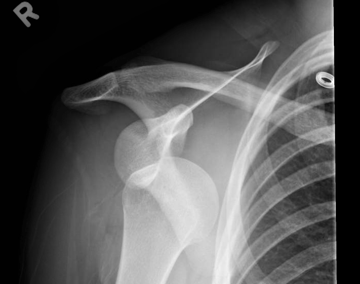
Ever had a case with a foreign body in the foot? Do you know how painful this is?! 22 yo patient sent in from clinic because he had accidentally stepped on a broken wine glass several days before. The clinic removed some of the glass a few days before but returned because of ongoing pain. Here is his x-ray from the clinic.


In this case the glass is radio-opaque with a few pieces present. The patient wanted to be knocked out for this  because of the amount of pain trying to remove the glass last time. The first task is to locate the glass. Seem pretty easy, right?


 The glass FB on the still image is hyperechoic and causes a shadow and/or reverberation artifact. It has been highlighted on the still image, but watch the video...


A step off pad would have enhanced the visualization and detection of the foreign body. This can be done simply by using about 1 cm thickness of gel and suspending the transducer in the gel above the area of interest or getting a mini-bag of saline and scanning through that.

Next step ... removal...

Local anesthetic in this area is still extremely painful. Other options include procedural sedation and analgesia or a local block. These can be done blind, but studies have shown more effective blocks under direct visualization with ultrasound are more effective ...


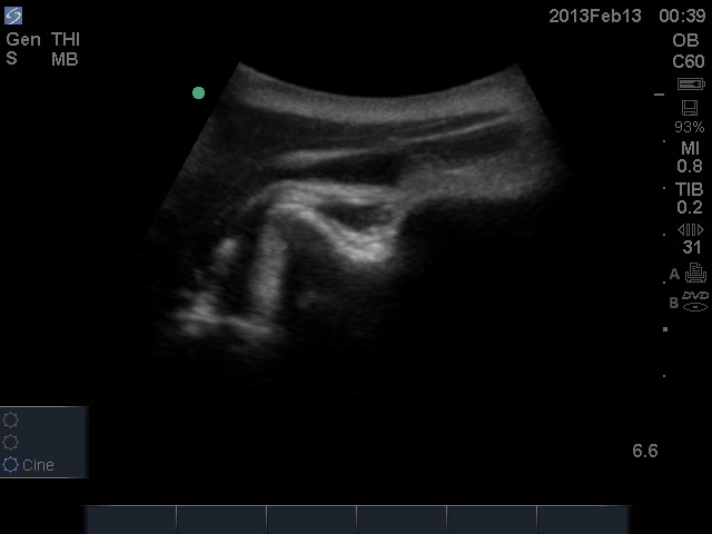
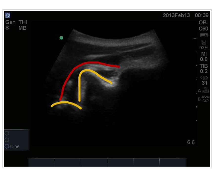
The posterior tibial nerve covers the heel completely so all you need to do is block this nerve as opposed to a complete ankle block. The anatomy is shown above. The transducer is placed just above the medial malleolus and vessels and the PTN can be visualized. 

Informed consent must be obtained and should include that lidocaine will last about 5-8 hours. Lidocaine WITHOUT epinephrine is used. During this time NO weight bearing should occur for the risk of stepping on something else as well as the risk of falls. Comparisons can be made with dental freezing and no chewing because the risk of gnawing off your cheek!! All patients are discharged with crutches. 


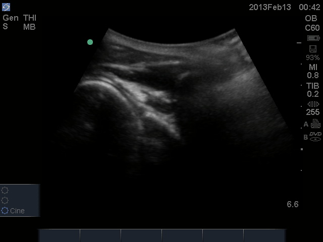
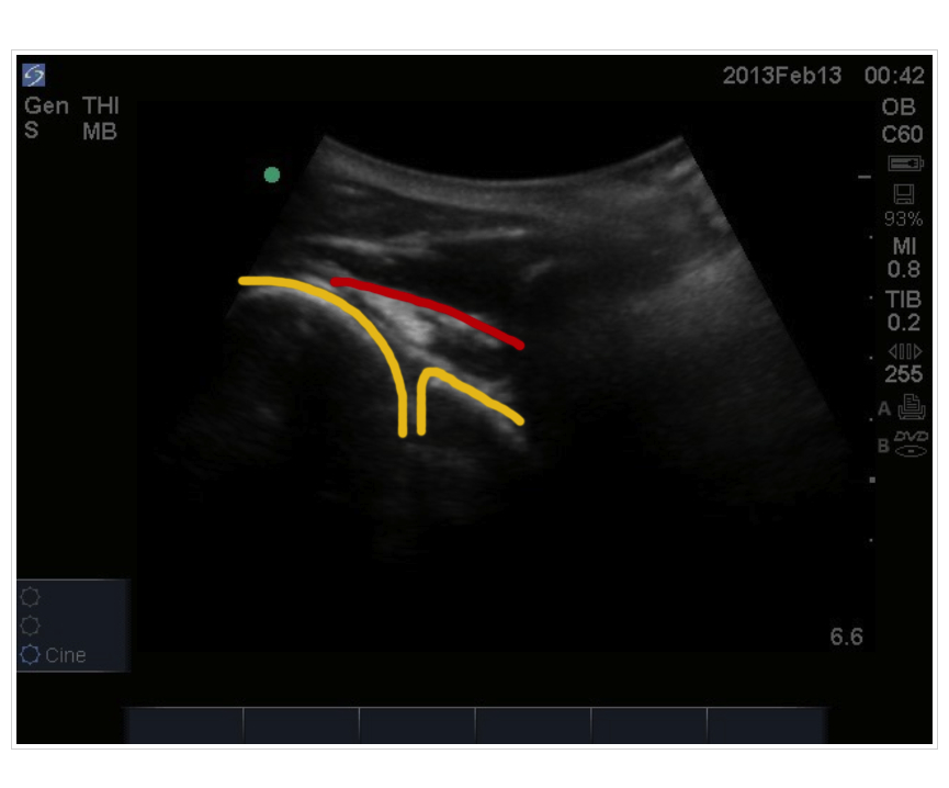
In the cine clip above, Dr. Vakani (our very own emerg resident) has advanced the needle towards the PTN avoiding the vessels. The needle can be seen coming from the left of the screen.


You can see now that the PTN is easily visualized by its honeycomb appearance as it is bathed around with lidocaine. As the needle is withdrawn, lidocaine is seen injected just above the fascia which would have resulted in a poor block if this was the only area where the lidocaine was injected.

Piece of cake? ... Not so fast .... Even though you know where the foreign body is AND you have great analgesia ... searching for these is still like a needle in the haystack. Give yourself a time limit ... 30 minutes maximum and consider calling in a consultant if it takes more time than this to do.


Hooray!! Everyone's happy right? .....

Nothing is 100% ... knowing that the foreign body in this case is radio-opaque the patient was sent back for repeat x-rays which demonstrated residual glass in the heel. A repeat ultrasound may have less utility because with dissection air is often introduced which makes the accuracy of ultrasound to pick up foreign bodies limited.  The patient was subsequently referred to orthopedics who then removed the remaining foreign bodies.

In summary:

1. Consider ultrasound guided regional nerve blocks for analgesia
2. Be sure to advise of all the risks and duration of the block to the patient
3. When removing foreign bodies:

  • give yourself a time limit
  • you may not get everything out even though you think you have (advise the patient accordingly before you start)
Great resources:


Thank you to the patient who kindly consented for us to put this case up as well as Dr. Vakani for letting me tag along! Please feel free to provide any feedback.

Yeah ultrasound!!


Monday 22 July 2013

Annual Intro Course a success!

The third week of July is always a big week on the Ottawa PoCUS calendar, as we hold our annual intro course for our new R1s and CCFP(EM) residents. 

We had a good time!


The ancient master passes on his skills...


Apprentice instructor JC gets enthusiastic....


This year's ultrasound fellow, Mark Taylor, was a hit!


Apprentice instructor Mike shows off some comet tails...

Next week, the ultrasound team returns with the procedural course as part of the Sim Boot Camp!

Friday 14 June 2013

Hey Folks!

Last month I took part in the Point of Care Ultrasound Interest Group in University of Western Ontario in London, Ontario. This was via a google hangout with Dr. Arntfield and Dr. Thompson. Thanks to Chris Byrne for hosting. The topic was Ultrasound in Medical Education and students had an opportunity to submit questions during the live broadcast. You can check out the recording here

Michael


Friday 24 May 2013

Journal club May 29, 2013

Intraoral and transcutaneous cervical ultrasound in the differential diagnosis of peritonsillar cellulitis and abscesses.

Bernardo Cunha Araujo Filho, Flavio A. Sakae, Luiz Ubirajara Sennes, Rui Imamura, Marcus R. de

BRAZILIAN JOURNAL OF OTORHINOLARYNGOLOGY 72 (3) MAY/JUNE 2006

Journal club goes international this month - see you there!

Friday 17 May 2013

Hong Kong - Shenzhen EM Conference

This post comes a bit late after my return, but things are fast and furious! The sabbatical ended with participation in the Hong Kong - Shenzhen EM Conference. This was a fantastic experience and introduced me to the challenges of communicating with physicians where English is a second or third language. Ever heard of Shen-Fu Injections for post-cardiac arrest syndromes! I certainly had not until this conference. By communicating and reading only in English -- are we missing out what the rest of the world has to offer?


Thursday 18 April 2013

Greetings now from Singapore (well a week late). This has been one of the highlights of the trip. By taking part in the Society for Emergency Medicine in Singapore (SEMS) Annual Scientific Meeting, I met many physicians from around South East Asia and beyond. By listening and speaking to such a diverse crowd at varying stages in the practice of Emergency Medicine, I have been able to gain a better appreciation of Emergency Medicine in general. The conference was a well organized, high quality conference which I came away with some educational pearls. In addition, taking part in the post-conference Winfocus sessions afterwards afforded me the opportunity to interact with other ultrasonography keeners. Great forum to exchange ideas and approaches to ultrasonography. The Korean dinner was real yummy too!
Winfocus Team post conference SEMS 2013
 
The conference and Winfocus course both took place in the beautiful surroundings of Khoo Teck Puat Hospital in Singapore. This is a new a great new facility with it's own lake for peaceful walks and tranquility steps away from the emergency department. Yes, that really is the waiting board -- 311 visits at 18:00 starting from 24:00 and only a 72 minute wait! Wow!

Wednesday 3 April 2013

Journal Club April 3, 2013

Use of bedside sonography for diagnosing acute epiglottitis in the emergency department: A preliminary study. J Ultrasound Med 2012; 31; 19-22.

Saturday 23 March 2013

Dr. Michael Woo and Dr. Gaby Blecher
Another update from Down Under...I had the opportunity to spend the day with Dr. Gaby Blecher and the Emergency Registrars at Monash Medical Centre in Melbourne, Australia. It was wonderful to catch up with Dr. Blecher who did a research fellowship at UOttawa with Dr. Stiell and completed his thesis on Point-of-care ultrasonography for DVTs in ED patients. He is now incorporating PoCUS as part of clinical care for EPs at Monash Medical Centre. I was able to present the role of PoCUS in patients presenting with shortness of breath and take part in their journal club. Taking part in their POCUS meeting was enlightening as they have advanced their QA system and have strong collaborations with Radiology. This was an enthusiastic group with lots of ideas. Looking forward to sharing these ideas with you all and developing future collaborations. (Oh yeah, I have enjoyed the surf, sun, and food as well! :)

Wednesday 20 March 2013

Pericardial Effusion - Seen one lately?

A 60 year old man presents with shortness of breath on exertion.  You work him up, but chest x-ray, spirometry, ECG, and labs (troponin and D-dimer) are all normal.  Send him home, I guess...

Have you considered occult pericardial effusion?  I have diagnosed about six unexpected pericardial effusions in the last year, and I have learned a few lessons:

1) this is not as rare a diagnosis as you think
2) patients can look really good, even with impending tamponade

This is an easy diagnosis to miss, but in 2013, I don't think that's acceptable anymore.  Any well-trained emergency physician should be thinking about it and has a tool at hand to make the diagnosis quickly and cheaply.

The first video here is in the subxiphoid view.


The next video is the same patient, showing the effusion in the parasternal long.


The third video is from a different patient.  Notice here how the right ventricular wall is collapsing inward at end-diastole.  This is an echocardiograhic sign of tamponade.  For the clinician, tamponade should be a clinical diagnosis, but if you see this at the bedside, be worried!


Occult pericardial effusion is not hard to diagnose.  You just need to think of it, and be liberal in your use of PoCUS.  Unexplained dyspnea, weakness, hypotension, or tachycardia?  Get out your probe!

Wednesday 13 March 2013

Greetings from Melbourne, Australia.
One of the great things about academic emergency medicine is the opportunity to travel and meet other interesting emergency physicians. I had the opportunity to attend journal club with staff and emergency registrars as well as share a presentation on the role of PoCUS in patients presenting with shortness of breath. It was wonderful to share ideas and learn the different approaches to emergency care. The Alfred Hospital is the major trauma centre in Melbourne and a fantastic facility. Thank you to my hosts and I look forward to collaborating on projects in the future!

Trauma Bay with overhead plain film imaging. Spot the US machine!  


My hosts in Melbourne standing next to the Geiger counter :)

No need with US!

Monday 4 March 2013

EMUS Journal Club Wednesday March 6

9:15 am, Civic Small Conference Room

Sonographic Assessment of the optic nerve sheath in idiopathic intracranial hypertension
Jochen Bauerle, Max Nedelmann
J Neurol (2011) 258:2014-2019.

See you there!

Wednesday 27 February 2013

POCUS guided Shoulder Reduction

Check out these amazing images, graciously shared with us by Dr. Nicolas Chagnon of the Hopital Monfort!


US-guided closed reduction of a R anterior shoulder dislocation

Before (glenoid and humeral head in yellow, supraspinatus tendon in red)


After (glenoid and humeral head in yellow, supraspinatus tendon in red)


Posted 2013/02/15 @ 14:44 by N. Chagnon.

Tuesday 12 February 2013

Teaching POCUS at NCCEM!

Your ultrasound team was busy again on the weekend, teaching at two afternoon workshops during the 2013 National Capital Conference on Emergency Medicine.  Here are some shots of us teaching DVT scanning.


Sunday 27 January 2013

Journal Club, January 30, 2013

Emergency Thoracic Ultrasound in the Differentiation of the Etiology of Shortness of Breath (ETUDES): Sonographic B-lines and N-terminal Pro-brain-type Natriuretic Peptide in Diagnosing Congestive Heart Failure

Andrew S. Liteplo, MD, RDMS, Keith A. Marill, MD, Tomas Villen, MD, Robert M. Miller, MD, Alice F. Murray, MBChB, Peter E. Croft, BS, Roberta Capp, MD, and Vicki E. Noble, MD, RDMS

9:15am, Civic

Friday 25 January 2013

PoCUS saves hockey icon's wife

Would YOU have made the diagnosis? This happened at a community hospital.

http://www.thestar.com/news/gta/article/1319889

Time to think about the future and get the skills we all will need. Anyone can learn this. Paul Pageau is teaching advanced cardiac at NCCEM. Or take an EDE2 course!